
A corneal transplant, also known as a corneal graft, or as a penetrating keratoplasty, involves the removal of the central portion (called a button) of the diseased cornea and replacing it with a matched donor button of cornea. Corneal grafts are performed on patients with damaged or scarred corneas that prevent acceptable vision. This may be due to corneal scarring from disease or trauma. In this photo of a recent transplant, the fine V-shaped stitches can be seen clearly on the right side, though they extend all the way around the transplant.
A common indication for keratoplasty is keratoconus. The eye-care practitioner must decide when to recommend keratoplasty for the keratoconic patient. This is often not a simple, straight-forward decision. Keratoplasty for keratoconus is highly successful; however, there is a long recovery period and a risk of severe ocular complications. A number of factors must be considered in deciding when to do a keratoplasty. One of the most important is the patient’s functional vision. If the best acuity with their contact lenses prevents them from doing their job or carrying out their normal activities, a transplant must be considered. The actual measured visual acuity may be quite different for different patients. One patient may find that he/she can not do their job with 20/30 acuity while another patient may be very satisfied with 20/60 acuity.
Very careful contact lens fittings are necessary before recommending a corneal transplant. One study found that 69% of keratoconics, most referred for transplant, could be successfully fit with contact lenses if special lens designs were used. Thus, prior to transplant every effort should be made to optimally fit the patient with contact lenses, especially if there is not significant corneal scarring affecting vision. However, a few patients become intolerant to contact lenses, and require a transplant earlier than otherwise would be necessary. If the patient has a large area of thinning, a very decentered cone or significant blood vessel growth into the usually clear cornea, called neovascularization, a transplant may be performed earlier than otherwise indicated by the visual performance, as these factors may require a larger than normal transplant button size and/or increase the chance of rejection if allowed to advance too far.
The healing process following transplant is long, often taking a year or longer. The time from surgery to the removal of the stitches is commonly 6 to 17 months. The patient may be on steroids for months. Initially following surgery, the donor button is swollen and even following healing the button is usually thicker than the corneal bed in which it rests. The photo on the left shows a thick white ring of scarring between the donated button and the patient’s outer cornea. The white radial marks are where stitches were placed to hold the transplant in position.Graft rejection reactions occur in 11% to 18% of the patients . Signs of graft rejection include ciliary flush, anterior chamber flare, keratic precipitates, Khodaoust line and Krachmer’s spots. Signs of graft rejection are reported to occur from 1 month to 5 years following surgery . The rejection rate for bilateral grafts is higher than if only one eye is grafted. In the bilateral cases, when a rejection reaction occurs it is commonly in both eyes. If the second eye is to be grafted, there is usually a period of at least a year between grafts. If signs of rejection occur, aggressive treatment with steroids is begun. Usually the reaction is overcome and the graft remains clear. Over 90% of the corneal grafts are successful with some studies reporting 97% to 99% success rates at 5 and 10 years. Large amounts of astigmatism are common following keratoplasty. One study found an average of 5.56 DC with a range from 0 to 17 D following suture removal, with other studies showing an average of 5.4 DC and 4.5 DC.
Texas Eye Institute is proud to provide five convenient locations for your eye care needs. Visit one of our convenient locations in Angleton, Sugarland, Southwest Houston, Katy, or Southeast Houston to see why the Texas Eye Institute is the best choice to care for your vision. Need LASIK in Houston? What about a comprehensive eye exam in Sugarland? See our locations page to find our practice nearest you!
Our Beechnut location is relocating to a new home starting May 26, 2026.
Find us at our new address:
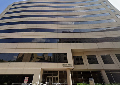
Memorial Hermann Medical Plaza 2
7737 Southwest Freeway, Houston, TX 77074
Clinic: Suite 600
Pediatrics: Suite 610
Optical: Suite 110
We’re excited to continue serving you in a bigger, better space. Same great care, in a new office nearby. Just behind our current Beechnut building.